Human Physiology / Autonomic nervous system
24 Sept Seminar 2
24 September 2026
1. What the ANS controls
The autonomic nervous system (ANS) controls things mostly automatically:
- Heart muscle: heart rate and strength of beating.
- Smooth muscle: in organs such as gut, bladder and blood vessels.
- Glands: saliva, sweat and other secretions.
Skeletal muscle, such as your arm muscles, uses somatic motor nerves, not the ANS.
- CNS: brain and spinal cord.
- PNS: nerves and ganglia outside them. Includes sensory pathways and somatic/autonomic motor pathways.
- Somatic is neither sympathetic nor parasympathetic. It is a separate motor division.
CNS/PNS tells you where. Somatic/autonomic tells you what kind of control. Both motor systems receive commands from the CNS.
2. The three autonomic divisions
- Sympathetic: prepares the body for action. Generally speeds the heart and reduces digestion.
- Parasympathetic: generally slows the heart and supports digestion and bladder emptying.
- Enteric: local nerve circuits in the gut; coordinate movement and secretion.
These are not three switches for the whole body. Different organs can respond differently; sympathetic nerves also work at rest.
| Feature | Sympathetic | Parasympathetic |
|---|---|---|
| Origin | T1-L2 spinal cord: thoracolumbar. | Brainstem + S2-S4: craniosacral. |
| Ganglion location | Usually closer to the spinal cord. | Near or inside the organ. |
| Typical axon lengths | Short first; long second. | Long first; short second. |
Vagus = cranial nerve X: major parasympathetic supply to organs in the chest and abdomen. It also carries sensory information back towards the brain.
3. How a nerve sends a message
A nerve releases a chemical called a neurotransmitter. It binds a receptor, a protein on the receiving cell.
Nerve → chemical message → receptor → cell responds
The response depends on which receptor and which cell receive the message.
- Acetylcholine (ACh) binds nicotinic or muscarinic receptors. Together, these are called cholinergic receptors.
- Noradrenaline (NA) and adrenaline bind alpha or beta receptors, with different strengths of action. These are adrenergic receptors.
The transmitter is the message. The receptor receives it. Nn, Nm, M2, M3, alpha and beta are receptors, not transmitters.
4. The two-neuron arrangement
Most autonomic commands pass through two separate neurons:
CNS → first neuron → ganglion → second neuron → organ
- First neuron = preganglionic. Its cell body is inside the CNS; its axon reaches the ganglion.
- Second neuron = postganglionic. Its cell body is inside the ganglion; its axon goes to the organ.
- Ganglion: a cluster of neuron cell bodies outside the CNS.
- Nucleus: a cluster of neuron cell bodies inside the CNS. Here, it does not mean the nucleus inside one cell.
The neurons do not merge. At the ganglion, the first releases ACh across a tiny gap to receptors on the second.
Both autonomic branches use ACh → Nn at their ganglia. There are many ganglia; "both" means sympathetic and parasympathetic, not just two ganglia.
5. The pathways, in order
Somatic → skeletal muscle
CNS → one neuron → ACh → Nm receptor on skeletal muscle → contraction
No autonomic ganglion.
Parasympathetic
CNS → first neuron → ACh → Nn on second neuron in ganglion → second neuron → ACh → muscarinic receptor on organ
Sympathetic: usual pathway
CNS → first neuron → ACh → Nn on second neuron in ganglion → second neuron → noradrenaline → alpha/beta receptor on organ
Adrenal medulla
CNS → first neuron → ACh → Nn on chromaffin cell → adrenaline + noradrenaline enter blood → alpha/beta receptors on organs
- Adrenal gland: whole organ above the kidney.
- Adrenal medulla: inner region of that gland.
- Chromaffin cells: cells in the medulla that secrete mainly adrenaline, plus some noradrenaline.
Why "usual" sympathetic pathway?
- Sweat glands: sympathetic nerves release ACh onto M3 receptors, not noradrenaline.
- Adrenal medulla: secretes hormones into blood instead of sending a second axon to each organ.
Pathway diagram
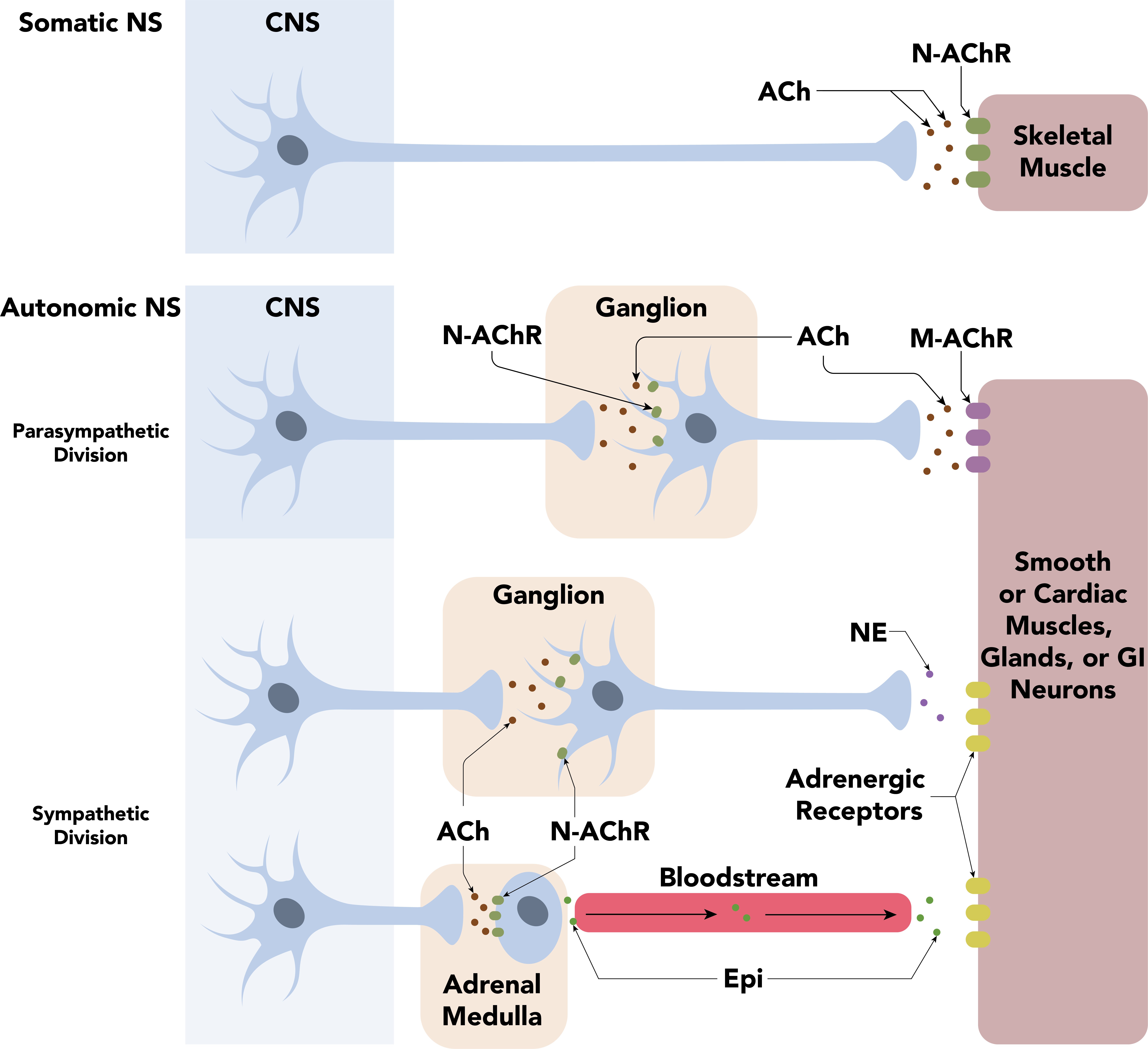
Daniel Walsh & Alan Sved · CC BY-SA 4.0.

6. The receptors worth knowing
| Receptor | Main thing to remember |
|---|---|
| Nn Neuronal nicotinic acetylcholine receptor | Receives ACh in sympathetic and parasympathetic ganglia, and on adrenal chromaffin cells. Excites the receiving cell. |
| Nm Muscle-type nicotinic acetylcholine receptor | Receives ACh on skeletal muscle → excitation leading to contraction. |
| M2 M2 muscarinic acetylcholine receptor | Slows the heart and conduction through the AV node. |
| M3 M3 muscarinic acetylcholine receptor | Makes glands secrete; contracts certain smooth muscles, including bladder wall and the muscle that constricts the pupil. |
| α1 Alpha-1 adrenergic receptor | Contracts blood-vessel smooth muscle → constriction. Contracts radial iris muscle → pupil dilation. |
| α2 Alpha-2 adrenergic receptor | On a nerve terminal: tells it to release less noradrenaline. |
| β1 Beta-1 adrenergic receptor | Makes the heart beat faster and stronger; stimulates renin release in the kidney. |
| β2 Beta-2 adrenergic receptor | Relaxes airway smooth muscle → airways widen. Can also relax some blood vessels. |
| β3 Beta-3 adrenergic receptor | Relaxes bladder wall → helps store urine. |
Numbers identify different subtypes, not steps in a sequence or levels of strength.
How receptors work
- Nicotinic: ACh binds → a channel in the receptor opens → ions move → the cell is electrically excited.
- Muscarinic and adrenergic: the receptor activates a G protein inside the cell, which changes enzymes or ion-channel activity. These are G-protein-coupled receptors, or GPCRs.
Same message, different response: ACh can excite a ganglion neuron through Nn, but slow the heart through M2. Always ask which receptor, on which cell?
7. Important organ examples
- Heart: sympathetic speeds it up; parasympathetic slows it.
- Gut: sympathetic generally reduces activity; parasympathetic generally increases it.
- Bladder storage: wall relaxed, outlet closed.
- Bladder emptying: wall contracts, outlet relaxes. Emptying is called voiding or micturition.
- Pupils: sympathetic widens them; parasympathetic narrows them.
The external urethral sphincter is skeletal muscle under somatic control; it must also relax for urination.
8. Three mechanisms to explain
- Adrenal medulla: ACh activates chromaffin cells → adrenaline/NA enter blood → reach many organs.
- Enteric reflex: gut contracts behind food and relaxes ahead, pushing it forward.
- Standing up: blood pressure briefly falls → pressure-sensor firing falls → brainstem increases sympathetic and reduces cardiac vagal activity → heart speeds up and vessels constrict → pressure recovers.
Afferent carries sensory information towards the CNS. Efferent carries commands away.
Negative feedback: the response opposes the original change. Falling pressure triggers changes that bring pressure back up.
9. One important drug example
Atropine blocks muscarinic receptors.
Therefore: less saliva and sweat, dilated pupils, usually faster heart.
It blocks sweating even though sweating is sympathetic, because the sweat gland uses ACh and muscarinic receptors.
It does not block the nicotinic Nn or Nm receptors.
10. Where does ACh come from?
The nerve ending makes it, stores it and releases it when an electrical signal arrives.
- The ending makes ACh from choline + acetyl-CoA.
- It stores ACh in tiny membrane bags called vesicles.
- An electrical impulse travels down the axon to the ending.
- Calcium channels open → calcium enters.
- Calcium triggers vesicles to fuse with the membrane → ACh is released.
- ACh crosses the gap and binds receptors on the next cell.
Electrical signal along the nerve → chemical signal across the gap.
- At a ganglion, the first neuron supplies ACh.
- At a parasympathetic target, the second neuron makes and releases its own ACh.
- The same ACh molecules do not travel along the entire pathway.
- Acetylcholinesterase breaks ACh down afterward. The ending takes choline back up and reuses it.
11. What is cAMP?
cAMP = cyclic adenosine monophosphate. It is a messenger inside the cell that helps pass on a signal received at the surface.
Adrenaline → beta receptor on the surface → Gs protein → enzyme makes cAMP → processes inside the cell change
- First messenger: the chemical arriving outside, e.g. adrenaline.
- Second messenger: cAMP passing the message inside.
- Gs increases cAMP. Gi decreases cAMP. These G proteins regulate the enzyme adenylyl cyclase.
- Gq uses another pathway, including release of stored calcium inside the cell.
In your receptor table: beta receptors use Gs; alpha-2/M2 use Gi; alpha-1/M3 use Gq.
The cell matters: increased cAMP can make the heart beat faster/stronger, but make airway smooth muscle relax.